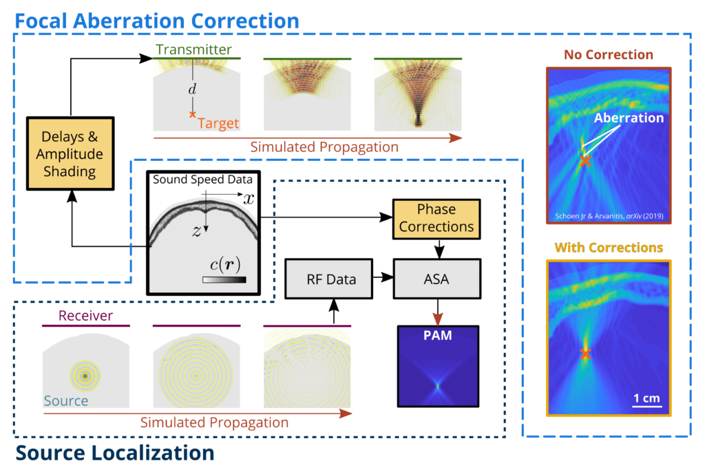
The complex acoustic environment presented by the skull induces significant aberration to ultrasound waves that makes trascranial focusing and imaging challenging. Our research in this area aims to establish computationally efficient methods that fully account for the skull induced aberrations and enable new therapeutic interventions and diagnostics.
Aberration Correction
While the angular spectrum approach affords very high intrinsic computational efficiency, as it operates in the frequency domain, its derivation does not account for heterogeneity of the medium. We have recently extended the angular spectrum approach for nonuniform media, including an approximate numerical method in the general case, and a closed-form analytical solution in the case of a stratified (layered) medium. Simulations showed that the general solution enables accurate (sub-millimeter) trans-skull focusing and point source localization that is orders of magnitude faster than simulation-based methods. Our approach provides an extremely efficient method for correcting skull-induced distortions. The enhanced capabilities of the proposed algorithm may enable new therapeutic interventions and build new and more efficient FUS systems.
| Flow of computations for focal aberration correction and improved source localization. All corrections are computed (yellow boxes) from the known sound speed variation c(r) (black box, center). In the case of focusing, the phase and amplitude of P due to a delta function at the desired focus and applied to the transmitting elements. In the case of PAM, the measured field P is propagated through the plane to form an intensity map from which the source location is extracted. [Schoen Jr & Arvanitis, IEEE T. Med. Imag. 39(5) (2020)] |  |
Image Guided Therapy
Despite its enormous potential for therapy, acoustic cavitation is also associated with increased risks, thus methods to monitor and map acoustic cavitation are of high importance in the effort to establish new therapeutic interventions. We recently demonstrated that the angular spectrum approach is able to localize microbubble activity at orders of magnitude faster (>1000 in 3D) than the established time domain methods, without compromising the spatial resolution or the signal to noise ratio, while retaining the information about the type of oscillation. By recognizing and underscoring the potential of the angular spectrum approach for real-time monitoring, imaging, and characterization of the inherently non-linear microbubble oscillations, our work, including the extended heterogeneous angular spectrum approach, is poised to transform the use of acoustic cavitation for treating human disease.
Computational Ultrasound
Diffraction places a fundamental limit on the spatial resolution of any imaging system. Our research explores the use of fast spectral methods, like the angular spectrum approach, for trans-skull sub-diffraction imaging of microbubbles. In addition to fast and spectrally resolved reconstructions we are interested in identifying computationally efficient peak finding algorithms and methods for automatic segmentation and characterization of vessel structure and flow velocities. We envision that these methods will enhance our ability to diagnose diseases that present aberrant microvascular structure and function (e.g. cancer) and assess therapies that promote vascular remodeling and regeneration.
 |
(Left) Spectrally-resolved super-resolution techniques allow recovery of velocity and structural information about the vessel from the recorded bubble emissions. (Right) As the angular spectrum approach and peak association methods extend naturally to higher dimensions, the image quality of 3D PAMs may be improved without significantly higher computational complexity. [Under Review]. |